Showing posts with label respiratory system. Show all posts
0
Download / swf / 330 Kb


This file will show you about anatomy of lung animation and disease of lung using swf format or flash media

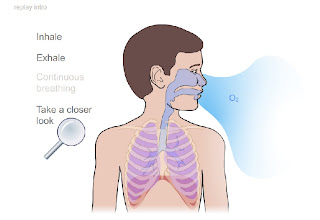
Lung is one of respiratory organ. The primary function of the respiratory system is to supply the blood with oxygen in order for the blood to deliver oxygen to all parts of the body. The respiratory system does this through breathing. When we breathe, we inhale oxygen and exhale carbon dioxide. This exchange of gases is the respiratory system's means of getting oxygen to the blood.

Respiration is achieved through the mouth, nose, trachea, lungs, and diaphragm. Oxygen enters the respiratory system through the mouth and the nose. The oxygen then passes through the larynx (where speech sounds are produced) and the trachea which is a tube that enters the chest cavity. In the chest cavity, the trachea splits into two smaller tubes called the bronchi. Each bronchus then divides again forming the bronchial tubes. The bronchial tubes lead directly into the lungs where they divide into many smaller tubes which connect to tiny sacs called alveoli. The average adult's lungs contain about 600 million of these spongy, air-filled sacs that are surrounded by capillaries. The inhaled oxygen passes into the alveoli and then diffuses through the capillaries into the arterial blood. Meanwhile, the waste-rich blood from the veins releases its carbon dioxide into the alveoli. The carbon dioxide follows the same path out of the lungs when you exhale.

The diaphragm's job is to help pump the carbon dioxide out of the lungs and pull the oxygen into the lungs. The diaphragm is a sheet of muscles that lies across the bottom of the chest cavity. As the diaphragm contracts and relaxes, breathing takes place. When the diaphragm contracts, oxygen is pulled into the lungs. When the diaphragm relaxes, carbon dioxide is pumped out of the lungs.
0

download / swf / 1181 Kb

The air finally ends up in the 600 million alveoli. As these millions of alveoli fill up with air, the lungs get bigger. Remember that experiment where you felt your lungs get larger? Well, you were really feeling the power of those awesome alveoli!

It's the alveoli that allow oxygen from the air to pass into your blood. All the cells in the body need oxygen every minute of the day. Oxygen passes through the walls of each alveolus into the tiny capillaries that surround it. The oxygen enters the blood in the tiny capillaries, hitching a ride on red blood cells and traveling through layers of blood vessels to the heart. The heart then sends the oxygenated (filled with oxygen) blood out to all the cells in the body.

When it's time to exhale (breathe out), everything happens in reverse: Now it's the diaphragm's turn to say, "Move it!" Your diaphragm relaxes and moves up, pushing air out of the lungs. Your rib muscles become relaxed, and your ribs move in again, creating a smaller space in your chest.

By now your cells have used the oxygen they need, and your blood is carrying carbon dioxide and other wastes that must leave your body. The blood comes back through the capillaries and the wastes enter the alveoli. Then you breathe them out in the reverse order of how they came in — the air goes through the bronchioles, out the bronchi, out the trachea, and finally out through your mouth and nose.

The air that you breathe out not only contains wastes and carbon dioxide, but it's warm, too! As air travels through your body, it picks up heat along the way. You can feel this heat by putting your hand in front of your mouth or nose as you breathe out. What is the temperature of the air that comes out of your mouth or nose?

With all this movement, you might be wondering why things don't get stuck as the lungs fill and empty! Luckily, your lungs are covered by two really slick special layers called pleural (say: ploo-ral) membranes. These membranes are separated by a fluid that allows them to slide around easily while you inhale and exhale.
0

download / swf / 79 Kb
download 2

Breathing is so vital to life that it happens automatically. Each day, you breathe about 20,000 times, and by the time you're 70 years old, you'll have taken at least 600 million breaths.

All of this breathing couldn't happen without the respiratory system, which includes the nose, throat, voice box, windpipe, and lungs.

At the top of the respiratory system, the nostrils (also called nares) act as the air intake, bringing air into the nose, where it's warmed and humidified. Tiny hairs called cilia protect the nasal passageways and other parts of the respiratory tract, filtering out dust and other particles that enter the nose through the breathed air.

Air can also be taken in through the mouth. These two openings of the airway (the nasal cavity and the mouth) meet at the pharynx, or throat, at the back of the nose and mouth. The pharynx is part of the digestive system as well as the respiratory system because it carries both food and air. At the bottom of the pharynx, this pathway divides in two, one for food (the esophagus, which leads to the stomach) and the other for air. The epiglottis, a small flap of tissue, covers the air-only passage when we swallow, keeping food and liquid from going into the lungs.

The larynx, or voice box, is the uppermost part of the air-only pipe. This short tube contains a pair of vocal cords, which vibrate to make sounds.

The trachea, or windpipe, extends downward from the base of the larynx. It lies partly in the neck and partly in the chest cavity. The walls of the trachea are strengthened by stiff rings of cartilage to keep it open. The trachea is also lined with cilia, which sweep fluids and foreign particles out of the airway so that they stay out of the lungs.
0

download / swf / 22 Kb
download 2

From the outside, lungs are pink and a bit squishy, like a sponge. But the inside contains the real lowdown on the lungs! At the bottom of the trachea (say: tray-kee-uh), or windpipe, there are two large tubes. These tubes are called the main stem bronchi (say: brong-kye), and one heads left into the left lung, while the other heads right into the right lung.

Each main stem bronchus (say: brong-kuss) — the name for just one of the bronchi — then branches off into tubes, or bronchi, that get smaller and even smaller still, like branches on a big tree. The tiniest tubes are called bronchioles (say: brong-kee-oles), and there are about 30,000 of them in each lung. Each bronchiole is about the same thickness as a hair.

At the end of each bronchiole is a special area that leads into clumps of teeny tiny air sacs called alveoli (say: al-vee-oh-lie). There are about 600 million alveoli in your lungs and if you stretched them out, they would cover an entire tennis court. Now that's a load of alveoli! Each alveolus (say: al-vee-oh-luss) — what we call just one of the alveoli — has a mesh-like covering of very small blood vessels called capillaries (say: cap-ill-er-ees). These capillaries are so tiny that the cells in your blood need to line up single file just to march through them.
0

download / swf / 99 Kb

Breathing is defined as "the exchange of gases between the cells of an organism and the external environment" by Kendall and Kendall in their classic physical therapy text, Muscles - Testing and Function.

While it is true that the physiology of breathing is complex and extensive, the process of getting those gases from the environment to the cellular level of the body can be observed and understood by the general, non-medical population. Understanding the mechanics of breathing can facilitate the doing of breathing exercises for general relaxation, pain management, general health promotion and the increase of energy.
0
download  / swf / 238 Kb


In humans and mammals, respiratory gas exchange is carried out by mechanisms of the heart and lungs. Ventilation is the process of air movement into and out of the lungs. Once air enters the lungs, diffusion of O2 and CO2 occurs in the alveoli. The oxygenated blood is then perfused throughout the body where gas exchange occurs in the capillary beds.[1] The blood is subjected to a transient electric field (QRS waves of the EKG) in the heart, which dissociates molecules of different charge. The blood, being a polar fluid, aligns dipoles with the electric field, is released, and then oscillates in a damped driven oscillation to form Y or Osborn Waves, V, U, and Y waves. The electric field exposure and subsequent damped driven oscillation dissociate gas from hemoglobin, primarily CO2, but more important, BPG, which has a higher affinity for hemoglobin than does oxygen, due in part to its opposite charge. Completely-dissociated hemoglobin (which will even effervesce if the electric field is too strong — the reason defibrillation joules are limited, to avoid bubble emboli that may clog vessels in the lung) enters the lung in red blood cells ready to be oxygenated.

The primary force applied in the respiratory tract is supplied by atmospheric pressure. Total atmospheric pressure at sea level is 760 mmHg (101 kPa), with oxygen (O2) providing a partial pressure (pO2) of 160 mmHg (21 kPa), 21% by volume, at the entrance of the nares, a partial pressure of 150 mmHg (20 kPa) in the trachea due to the effect of partial pressure of water vapor, and an estimated pO2 of 100 mmHg (13 kPa) in the alveoli sac, pressure drop due to conduction loss as oxygen travels along the transport passageway. Atmospheric pressure decreases as altitude increases, making effective breathing more difficult at higher altitudes. Higher BPG levels in the blood are also seen at higher elevations, as well.

In similar manner, CO2, which is a result of tissue cellular respiration, is also exchanged. The pCO2 changes from 45 to 40 mmHg (6.0 to 5.3 kPa) in the alveoli. The concentration of this gas in the breath can be measured using a capnograph. As a secondary measurement, respiration rate can be derived from a CO2 breath waveform.

Gas exchange occurs only at pulmonary and systemic capillary beds, but anyone can perform simple experiments with electrodes in blood on the bench-top to observe electric field-stimulated effervescence. Trace gases present in breath at levels lower than a part per million are ammonia, acetone, isoprene. These can be measured using selected ion flow tube mass spectrometry.
0
download / swf / 243 Kb

The function of the respiratory system is to bring oxygen to the blood and to remove the carbon dioxide. The respiratory system is composed of two parts:
  •    Conducting Portion
  •    Respiratory Portion

The Conducting Portion consists of a series of cavities and tubes conducting air to the lungs.
The Conducting Portion is composed of :

  •     Nose
  •     Nasopharynx
  •     Larynx
  •     Trachea
  •     Bronchi
  •     Bronchioles (terminal and respiratory).

Some of these structures lie outside the lungs (extrapulmonary) and need cartilaginous supports in their walls, which provide rigidity and flexibility. Some of the structures are inside the lungs (intrapulmonary), where the need for structural support is less. The respiratory bronchioles constitute an area of transition between the conducting and respiratory portions.

The Respiratory Portion consists of :
  •     Alveolar ducts
  •     Alveolar sacs
  •     Alveoli

The exchanges of gases (respiration) only occurs in the alveoli.
0


download / swf / 1181Kb
download 2

The lung (adjectival form: pulmonary) is the essential respiration organ in many air-breathing animals, including most tetrapods, a few fish and a few snails. In mammals and the more complex life forms, the two lungs are located near the backbone on either side of the heart. Their principal function is to transport oxygen from the atmosphere into the bloodstream, and to release carbon dioxide from the bloodstream into the atmosphere. This exchange of gases is accomplished in the mosaic of specialized cells that form millions of tiny, exceptionally thin-walled air sacs called alveoli.

To completely explain the anatomy of the lungs, it is necessary to discuss the passage of air through the mouth to the alveoli. Once air progresses through the mouth or nose, it travels through the oropharynx, nasopharynx, the larynx, the trachea, and a progressively subdividing system of bronchi and bronchioles until it finally reaches the alveoli where the gas exchange of carbon dioxide and oxygen takes place.

The drawing and expulsion of air (ventilation) is driven by muscular action; in early tetrapods, air was driven into the lungs by the pharyngeal muscles via buccal pumping, whereas in reptiles, birds and mammals a more complicated musculoskeletal system is used.

Medical terms related to the lung often begin with pulmo-, from the Latin pulmonarius ("of the lungs"), or with pneumo- (from Greek πνεύμων "lung").
0